
The Case of the Painful Knee: When the Clue Was Not in the Knee
Disclaimer: This blog is for general educational purposes only. It is not a diagnosis and does not replace advice from your dentist, oral surgeon, GP, or other healthcare provider. Every case is different, and treatment results vary from person to person. If you have severe swelling, fever, worsening pain, difficulty swallowing, difficulty breathing, bleeding, or signs of infection after dental surgery, please seek urgent medical or dental advice.A patient recently came to see me with discomfort around their left knee.
At first, the case seemed straightforward. They noticed some discomfort when they began jogging on a treadmill. However, after a short warm-up, the sensation disappeared and they could continue running for four to five kilometres without significant pain.
That detail did not immediately suggest a serious local knee injury.
But then came the unusual clue.
Later that night, while resting in bed, the knee became sore and achy. Even the light weight of the quilt resting over the leg could aggravate the area.
They could tolerate several kilometres of running, yet gentle pressure from a quilt caused discomfort.
Something did not quite add up.
Clue One: A Restricted Kneecap
Every clinical investigation needs to begin somewhere, so I started with the knee itself.
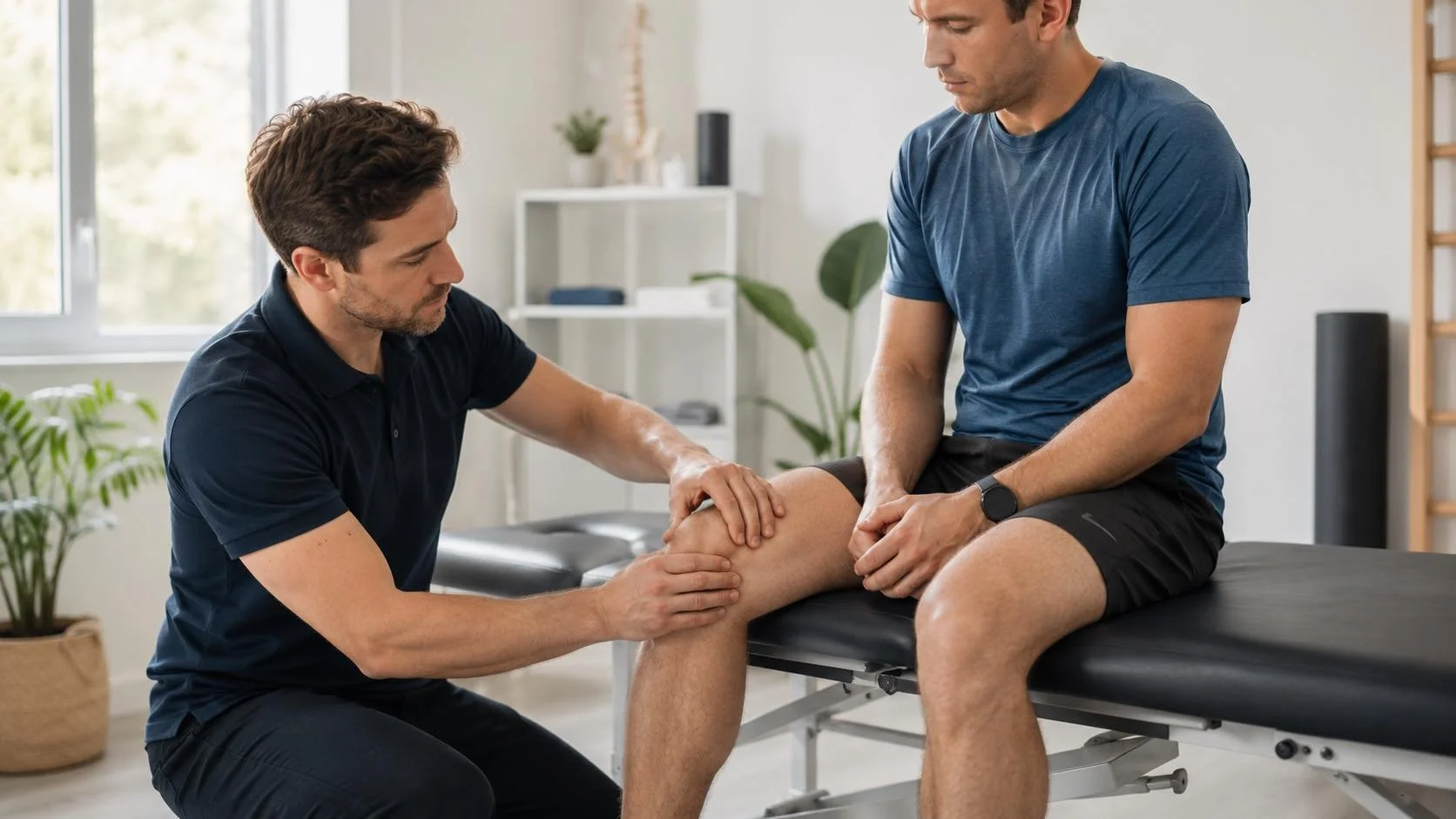
A basic orthopaedic assessment showed that the left kneecap, also known as the patella, was not moving as freely as the right.
There was also increased tension through the left quadriceps muscles.
This finding could easily lead to a familiar conclusion:
The quadriceps were tight, the kneecap was restricted, and the patient probably needed stretching, strengthening or running retraining.
Those approaches may still be useful.
However, they did not answer the most important question:
Why had the kneecap become restricted in the first place?
The restricted patella was a clue, but it might not have been the origin of the problem.
Clue Two: The Knee Was Not Giving the Whole Story
Using the Fascial Counterstrain assessment process, I began looking beyond the painful area.
The body first directed the assessment toward the junction between the neck and upper back, known as the cervicothoracic junction.
This region was significantly restricted.
That was an unexpected finding in someone presenting with knee discomfort.
However, in a clinical investigation, an unexpected clue should not be ignored simply because it is far away from the painful area.
Further assessment then led to the upper cervical spine.
This appeared to be an important part of the overall restriction pattern. I began by treating selected ligamentous and periosteal tissues around the upper neck.
The periosteum is the connective tissue covering the outer surface of a bone.
Once the upper neck restriction had improved, I reassessed the body to see where the next clue would lead.
Following the Trail Down the Body
The assessment returned to the cervicothoracic junction and then led to tissues associated with the suspension and support of the spinal cord.
From there, the next area was the dura.
The dura is a strong layer of connective tissue surrounding the brain and spinal cord. It extends from inside the skull, along the spinal canal and toward the lower end of the spine.
In other words, this tissue forms a continuous protective covering around much of the central nervous system.
The next clue appeared in the pelvic region.
Initially, I wondered whether the pelvis itself was misaligned or mechanically restricted. However, the assessment did not primarily indicate a joint alignment problem. Instead, it directed attention toward tissues associated with the parasympathetic nervous system.
After treating these areas, the assessment continued down the leg and eventually led to the Achilles tendon.
At this stage, the investigation had travelled from the upper neck, through the spinal tissues and pelvic region, and down toward the ankle.
The knee appeared to be part of a much larger pattern.
Returning to the Scene of the Pain
After addressing the initial restrictions, I returned to the knee and reassessed the patella.
Its mobility had improved significantly.
However, it was still not completely equal to the unaffected side.
The case was not solved yet.
Further assessment revealed tension associated with the left femur, or thigh bone. The body directed treatment toward the periosteal tissues surrounding the femur before progressing into the front muscular and fascial chain of the leg.
Once those areas had been treated, I returned to the knee again.
This time, the result was much clearer.
By the end of the 30-minute session:
the left patella moved as freely as the right
the quadriceps tension had settled
gentle jogging no longer reproduced the initial discomfort
The restricted kneecap had returned to normal mobility without treating the knee in isolation.
Was the Case Completely Solved?
Not yet.
The immediate findings were encouraging, but this was only a 30-minute treatment session.
The true test will be how the knee responds during the patient’s next longer run and whether the night-time aching returns.
I am awaiting their feedback at the next appointment.
Good clinical reasoning does not mean declaring victory after one session. It means observing the immediate change, testing the findings and continuing to monitor the patient’s response over time.
The Obvious Suspect: Weakness or Overuse
If I had followed a more conventional or local approach, the likely suspects might have included:
patellofemoral joint irritation
quadriceps weakness
gluteal weakness
running technique
training load
footwear
early joint degeneration
These possibilities are not necessarily wrong.
Exercise rehabilitation, strengthening and running assessment can all play an important role in treating knee pain.
However, in this case, they did not fully explain the findings.
The patient was already capable of running four to five kilometres. Their main physical restriction was poor patellar mobility, and that mobility changed after treating tissues located well away from the knee.
Had I immediately prescribed more strengthening without investigating further, I might have missed the restriction pattern maintaining the kneecap tension.
Strength Is Important, but It Is Not the Only Clue
It is tempting to believe that every painful joint simply needs stronger muscles around it.
But if strength alone prevented pain, everyone who regularly trained at the gym would live completely pain-free.
Clearly, that is not the case.
A person can be strong and still have:
restricted joint mobility
protective muscle tension
altered nervous-system sensitivity
connective-tissue restriction
poor load distribution
compensation from another part of the body
Strengthening may be part of the solution, but it should not automatically be assumed to be the whole solution.
The Main Lesson: Assessment Finds Hidden Restrictions
The painful area is always important.
However, it may be only the location where the body is displaying the final symptom.
A thorough assessment can help investigate:
whether the local joint is moving normally
whether nearby muscles are guarding
whether tension is being transmitted from another region
whether the nervous system is contributing to sensitivity
whether the restriction changes after treating another area
whether exercise should begin immediately or follow hands-on treatment
For this patient, the most useful clue was the restricted kneecap.
The most interesting finding was that the kneecap regained normal movement only after a broader pattern of restrictions had been addressed.
The knee was where the symptoms appeared.
The investigation suggested that the story was much bigger than the knee itself.
A Detective Approach to Knee Pain
Clinical assessment can be a little like detective work.
The first symptom is only the opening clue.
The physiotherapist then needs to ask:
What movements are restricted?
What makes the symptoms better or worse?
Does the painful area explain the full pattern?
Are other parts of the body contributing?
Does treating one area change the findings somewhere else?
Sometimes the final answer is found at the painful joint.
Sometimes the most important clue is somewhere unexpected.
That is why treatment should be guided by assessment rather than assumption.
Every patient is different, and no treatment can guarantee a particular outcome. Persistent or worsening knee pain should be assessed carefully, particularly when there is swelling, locking, instability, significant trauma, fever or difficulty bearing weight.
When knee discomfort continues despite rest, exercise or local treatment, a broader assessment may help uncover restrictions that have previously been missed.
Frequently Asked Questions
Can knee pain come from somewhere else in the body?
The painful sensation is experienced at the knee, but movement and tension elsewhere in the body may influence how forces are distributed through the leg. The hip, ankle, foot and spine may all contribute to the overall pattern.
Does knee pain always mean arthritis or degeneration?
No. Knee pain may arise from many different tissues and movement patterns. Symptoms alone cannot confirm degeneration. A clinical assessment, and sometimes medical imaging, may be needed when arthritis or another structural condition is suspected.
Why might a knee tolerate running but hurt under a quilt?
Pain sensitivity does not always correspond directly with the amount of load placed on a joint. Resting position, local tissue sensitivity, nerve irritation and protective muscle tension may all influence symptoms.
Should I stop running when I have knee discomfort?
Not necessarily. It depends on the severity of the symptoms, whether they worsen during or after running and whether there is swelling, instability or altered movement. Temporarily reducing distance or intensity may be appropriate until the problem has been assessed.
Is strengthening still important for knee pain?
Yes. Strengthening can improve capacity, control and confidence. However, exercise should ideally be based on the findings of the assessment rather than prescribed automatically for every knee problem.
What is Fascial Counterstrain?
Fascial Counterstrain is a gentle, hands-on assessment and treatment approach. It uses specific tender-point and tissue-response findings to identify areas of dysfunction across different body systems. Treatment is performed in comfortable positions without forceful manipulation.
If you are experiencing knee pain:
Experiencing persistent knee discomfort despite strengthening, rest or local treatment? A detailed physiotherapy assessment may help identify hidden restrictions affecting your movement. Contact Fascial Release Clinic in Glenelg to arrange an appointment.